
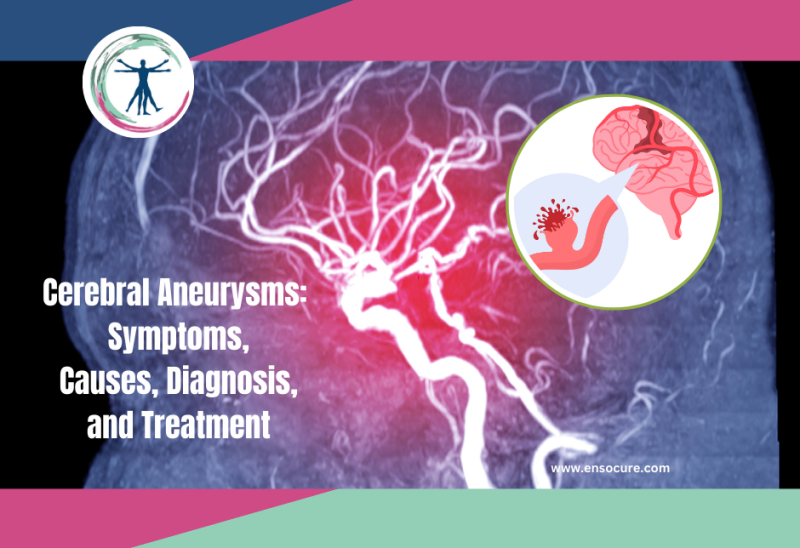
An aneurysm refers to a dilation or bulge in an artery, which is responsible for transporting oxygenated blood from the heart to different parts of the body. Weakened artery walls are at risk of developing aneurysms with the main risk factors being issues like high blood pressure and congenital abnormalities While most aneurysms develop in the aorta—the principal artery linking the heart to the chest and abdomen—they can occur in any artery. When aneurysms affect arteries in the brain, they are called cerebral aneurysms or intercranial aneurysms which can lead to potentially serious complications such as brain hemorrhage and stroke.
Prevalence
Although aneurysms can affect individuals of all genders and ages, certain types are more commonly observed in specific populations. In India intercranial aneurysms are regarded an incidental disease with not much data for specific prevalence. Research in North India found the incidence of aneurysms was 1.6% in females and 0.8% in males all of which were saccular. Further research has found the prevalence of Aneurysmal subarachnoid hemorrhage (SAH) in India varies from 0.75 to 10.3%. 40% of cerebral aneurysms were fatal. The biggest risk factors in India were found to be smoking and hypertension.

Symptoms of aneurysms vary depending on their severity and location. Small cerebral aneurysms do not rupture, but in severe cases, the artery might, resulting in internal bleeding, which can be life-threatening. Fortunately, there are treatment options available for this condition. It is important to understand all cerebral aneurysms have the potential to rupture and cause cerebral hemorrhage which is why a timely diagnosis and treatment is necessary.
Types of Cerebral Aneurysms
Currently, cerebral aneurysms are typically categorized into three primary types: Saccular, Fusiform and Mycotic
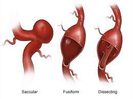
- Saccular Aneurysm: A saccular aneurysm develops as a rounded sac filled with blood, which is connected to a primary artery or one of its branches. Also referred to as a berry aneurysm due to its resemblance to a hanging berry, this type is the most common form of cerebral aneurysm. Cerebral aneurysms ate usually located on arteries situated at the base of the brain.
- Fusiform Aneurysm: A fusiform aneurysm protrudes or swells out uniformly on all sides of the artery.
- Mycotic Aneurysm: Mycotic aneurysms result from infections that can affect the arteries in the brain. These infections weaken the artery walls, leading to the formation of a bulging aneurysm.
Classification by Size
- Small aneurysms: Less than 11 mm in diameter
- Large aneurysms: 11 to 25 mm in diameter
- Giant aneurysms: >25 mm in diameter
Symptoms
Symptoms of a cerebral aneurysm can vary depending on whether the aneurysm has ruptured or not. However, common symptoms may include:
Unruptured Aneurysm
- Pain behind or above the eye
- Numbness and Weakness
- One side facial paralysis
- Dilated pupils
- Vision changes or double vision.
Ruptured Aneurysm
- Sudden and severe headache (like thunderclap headaches often described as the worst headache of one’s life)
- Nausea and vomiting
- Stiff neck
- Blurred or double vision
- Sensitivity to light
- Seizures
- Loss of consciousness
- Confusion or difficulty concentrating
- Weakness or numbness, typically on one side of the face or body
- Difficulty speaking or understanding speech
- Drooping eyelid
- Dilated pupils
- Changes in mental status or alertness
- A sudden increase in blood pressure
- High risk of cardiac arrest
It’s important to note that some cerebral aneurysms may not cause any symptoms and are only discovered incidentally during imaging tests conducted for other reasons. If you or someone else experiences any of these symptoms, especially a sudden and severe headache, it’s crucial to seek immediate medical attention as a ruptured cerebral aneurysm can be life-threatening.
Leaking Aneurysms
A leaking aneurysm in one which can release a small quantity of blood into the brain, a condition known as a sentinel bleed. This minor leakage from an aneurysm can sometimes lead to sentinel or warning headaches days or weeks before a major rupture occurs. However, it’s important to note that only a minority of individuals experience a sentinel headache prior to a rupture.
If you encounter a sudden, intense headache, particularly when accompanied by other symptoms, you need to seek medical attention.
Risk Factors of Cerberal Aneurysms
Cerebral aneurysms develop because of weakened artery walls, especially in the branch points where arteries are weakest. Cerebral aneurysms can occur at any age with the most common ages being 30-60. Here are the major risk factors of cerebral aneurysms:
- Having a family history of cerebral aneurysms or certain genetic disorders that weaken blood vessels
- Certain medical conditions, such as polycystic kidney disease, Ehlers-Danlos syndrome, Marfan syndrome, and arteriovenous malformations (AVMs), can increase the risk of cerebral aneurysms.
- Untreated hypertension (high blood pressure)
- Cigarette smoking
- Drug abuse, especially cocaine or amphetamines, (These raise blood pressure dangerously) Intravenous drug abuse is a cause of infectious mycotic aneurysms
- Age over 40
- Head trauma
- Brain tumor
- Infection in the arterial wall (mycotic aneurysm)
- Atherosclerosis, a condition characterized by the buildup of plaque in the arteries, can weaken blood vessel walls and raise the risk of aneurysm formation.
Risk factors for ruptured cerebral aneurysm
- Not all CA’s rupture, it depends on size location and growth including risk factors that speed up the possibility of a rupture. Risk factors include:
- Smoking
- High blood pressure.
- Size (Larger aneurysms are most likely to rupture).
- Posterior communicating artery aneurysms (arteries in the back part of the brain) and possibly those on the anterior communicating artery (a single artery in the front of the brain) are at high risk of rupture.
- Growing aneurysms are at increased risk of rupture.
- Family history of aneurysm rupture suggests a higher risk of rupture
- Individuals who have previously experienced ruptured cerebral aneurysms
Diagnosing Cerebral Aneurysms
Most cerebral aneurysms go unnoticed. They are only detected by imaging tests for other conditions or when a person experiences severe thunderclap headaches indicative of a ruptured aneurysm. Procedures include:
Computed Tomography (CT) Scan: A CT scan of the head is often the initial imaging test performed to detect a cerebral aneurysm, especially in emergency situations such as suspected ruptures. It provides detailed cross-sectional images of the brain and blood vessels.
Magnetic Resonance Imaging (MRI): MRI uses powerful magnets and radio waves to produce detailed images of the brain and blood vessels. It can provide more detailed information about the size, location, and shape of cerebral aneurysms compared to CT scans.
Magnetic Resonance Angiography (MRA): MRA is a specialized MRI technique that specifically focuses on imaging the blood vessels. It can provide detailed images of the blood vessels in the brain, including any abnormalities such as aneurysms.
Cerebral Angiography: Cerebral angiography, also known as digital subtraction angiography (DSA), is considered the gold standard for diagnosing cerebral aneurysms. It involves injecting a contrast dye into the blood vessels and taking X-ray images to visualize the blood flow and identify any abnormalities, including aneurysms.
Cerebrospinal fluid (CSF) analysis. A CSF test measures chemicals in cerebrospinal fluid. The test is done through a spinal tap where a lumbar puncture is performed in the lumbar spine to draw out a small amount of CSF. If bleeding is detected, additional tests would be needed to identify the exact cause.
Treatment of cerebral aneurysms
The treatment options for cerebral aneurysms depend on various factors, including the size, location, shape, and risk of rupture of the aneurysm, as well as the patient’s overall health and preferences. Treatment may include:
Observation: For small, unruptured aneurysms that are not causing symptoms and are deemed to be at low risk of rupture, close monitoring with regular imaging studies may be recommended. This approach is often preferred for elderly patients or those with significant comorbidities who may be at higher risk for surgical interventions.
Medication: Certain medications may be prescribed to manage symptoms, prevent complications, or reduce the risk of aneurysm rupture. These may include blood pressure-lowering medications to reduce stress on the blood vessel walls and medications to prevent or treat conditions such as seizures or headaches. Other medications include:
- Antiseizure drugs (anticonvulsants) to prevent seizures associated with a ruptured aneurysm.
- Calcium channel-blocking drugs to reduce the risk of stroke by vasospasm.
Surgical Clipping: Surgical clipping involves making a small opening in the skull (craniotomy) to access the aneurysm directly. A metal clip is placed across the neck of the aneurysm to permanently seal it off from the bloodstream, preventing the risk of rupture. Surgical clipping may be recommended for larger or complex aneurysms, as well as for patients who are not suitable candidates for endovascular therapy. In general, aneurysms that are completely clipped do not recur.
Endovascular Therapy: Endovascular therapy, also known as minimally invasive or catheter-based treatment, involves accessing the blood vessels through a small incision in the groin and threading a catheter (thin, flexible tube) through the blood vessels to the site of the aneurysm. Several endovascular techniques may be used to treat cerebral aneurysms, including:
- Coiling: In coil embolization, platinum coils are inserted into the aneurysm to fill it and promote clotting, effectively sealing off the aneurysm from the bloodstream and reducing the risk of rupture. This may need more than one treatment because of the possibility of the aneurysm re-occurring.
- Flow Diversion: Flow diverters are devices or stents placed, in the artery to reduce blood flow into the aneurysm. Usually performed in large aneurysms where platinum coil embolization is not an option.
- Stent-Assisted Coiling: In stent-assisted coiling, a stent is placed across the neck of the aneurysm to provide support and prevent coil migration during coil embolization.
CSF shunt: A shunt may be surgically inserted in the brain following rupture to funnel cerebrospinal fluid from the brain to elsewhere in the body. This is especially if CSF buildup causes pressure in surrounding areas.
Rehabilitative therapy: Individuals with subarachnoid hemorrhage might need physical, speech, and occupational therapy post treatment to regain lost function and learning how to cope with disabilities.
Risk Prevention
To prevent the risk of a cerebral aneurysm rupture, individuals should abide by the following:
- Avoid smoking
- Avoid drug abuse, cocaine, and stimulant drugs
- Good blood pressure management
- Eat a healthy diet
- Regular monitoring
- Prevent straining and strenuous physical activity
- Manage constipation through fiber
- Manage stress
- Limit caffeine and alcohol
- Take your medications
Current research is going on to develop new models to specifically diagnose cerebral aneurysms using the presence of molecules that can indicate the risk of a brain aneurysm rupturing. This can be performed by imaging allowing people to avoid surgery and get treated before an aneurysm ruptures.
Overall, adopting a healthy lifestyle, managing underlying medical conditions, and abiding by recommended treatment are essential for preventing the rupture of cerebral aneurysms and reducing associated risks. Regular follow-up with your doctor is crucial for ongoing monitoring and management of cerebral aneurysms. If you or any member of your family require post-surgical care, therapy, or counselling for cerebral aneurysms, contact Ensocure Integrated Medicine for a specialized health care support plan.