
An electrocardiogram (ECG or EKG) is a crucial test to diagnose and monitor your heart health. A non-invasive test, it has revolutionized the way we understand the heart’s electrical activity, offering invaluable insights into cardiac conditions.
What is an ECG?
The heart’s electrical impulses govern the contraction and relaxation of its muscles, orchestrating the rhythmic heartbeat. The ECG captures these impulses, revealing patterns and anomalies translating them into a visual representation that healthcare professionals use to assess cardiac health.
The Procedure
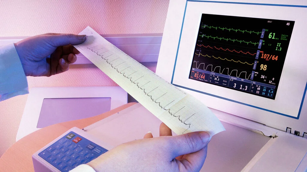
During an electrocardiogram, small electrodes are strategically placed on the patient’s chest, arms, and legs. These electrodes detect the electrical signals produced by the heart. The resulting data is then graphically represented on a monitor or paper, showcasing the heart’s electrical activity in the form of waves. Each wave corresponds to a specific phase of the cardiac cycle, and deviations from the norm can signal potential issues.
Parts of an ECG

The standard ECG has 12 leads. Six of the leads are considered “limb leads” because they are placed on the arms and/or legs of the individual. The other six leads are considered “precordial leads” because they are placed on the torso (precordium).
The six limb leads are called lead I, II, III, aVL, aVR and aVF. The letter “a” stands for “augmented,” as these leads are calculated as a combination of leads I, II and III.
The six precordial leads are called leads V1, V2, V3, V4, V5 and V6.
Common ECG Waves:
A normal ECG contains waves, intervals, segments and one complex, as defined below.
- P-Wave: Represents the electrical impulse through the atria or atrial depolarization
- QRS Complex: Consists of a Q wave, R wave and S wave and represents ventricular depolarization and contraction.
- T-Wave: Signifies ventricular repolarization and relaxation.
- Interval: The time between two specific ECG events. The intervals commonly measured on an ECG include the PR interval, QRS interval (also called QRS duration), QT interval and RR interval.
- Segment: The length between two specific points on an ECG that are supposed to be at the baseline amplitude (not negative or positive). The segments on an ECG include the PR segment, ST segment and TP segment.
- Point: There is only one point on an ECG termed the J point, which is where the QRS complex ends and the ST segment begins.
Understanding the normal patterns of these waves is crucial for interpreting ECG results accurately.
Why do you need an Electocardiogram
Your doctor may recommend an ECG to diagnose various conditions like:
Arrhythmias: ECG is highly effective in diagnosing irregular heart rhythms or arrhythmias. Whether it’s atrial fibrillation, bradycardia, or tachycardia, the distinct patterns on the ECG provide valuable information for the appropriate diagnosis and treatment.
Myocardial Infarction (Heart Attack): One of the primary uses of ECG is in detecting signs of a heart attack. Changes in the ST segment and the appearance of Q-waves can indicate myocardial damage, helping healthcare providers make swift decisions to save heart tissue.
Ischemia: ECG is a crucial tool for identifying insufficient blood supply to the heart muscle, known as ischemia. Changes in the ST segment during an ECG can reveal this condition, guiding healthcare professionals in managing and preventing further complications.
Heart Valve Disorders: Conditions affecting the heart valves, such as stenosis or regurgitation, can alter the normal flow of blood through the heart. ECG aids in identifying the impact of these disorders on the heart’s electrical activity.
Congenital Heart Abnormalities: ECG is instrumental in diagnosing congenital heart conditions, providing insights into irregularities in the heart’s structure and function from birth. Broken heart syndrome or Takotsubo Cardiomyopathy is another complex condition requiring an Electrocardiogram or ECG.
The Importance of Routine ECGs
While an Electrocardiogram is indispensable in diagnosing specific conditions, it is also used as a routine screening tool in various healthcare settings. Routine ECGs can help detect subtle abnormalities, enabling early intervention and preventive measures. They are particularly beneficial for individuals with risk factors like hypertension, diabetes, and a family history of heart disease. If your ECG tests are normal, you may not need further testing, but if they are not, if you have an irregularity that wasn’t detected, you might need to take another test called a holter monitor or further tests and treatments.